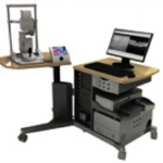
SPECTRALIS® OCT-Plus Imaging Platform
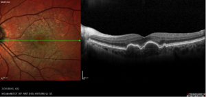 The SPECTRALIS® OCT Plus technology is a unique ophthalmic diagnostic imaging device used primarily to examine the structures of the retina, macula, and optic nerve. It combines two technologies, confocal scanning laser ophthalmoscopy (cSLO) and spectral domain optical coherence tomography (SD-OCT). It is the only spectral domain OCT imaging system to incorporate all the essential features listed below.
 We selected this technology for our practice because of the unique non-invasive screening capability for detecting eye disease using non-invasive ultra high quality visualization of the retinal structures using 3D- Ultra HD technology. Early detection of disease is critical for our patients and our primary concern and keeping your eyes healthy and preserving your best vision for years to come.
 Features:
- TruTrack™ active eye tracking: actively follows the patient’s eye during the scan, minimizing motion artifact and allowing targeted OCT scan placement.
- AutoRescan™: automatically places follow-up scans in precisely the same location as the baseline scan.
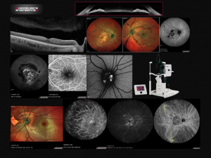
- FoDi™: fovea-to-disc alignment technology for more precise RNFL and ONH assessment.
- Heidelberg Noise Reduction™: combines multiple scans taken at the same location and eliminates noise from the image.
- Confocal Scanning Laser Ophthalmoscope: for high resolution fundus imaging.
- BluePeak™ with RegionFinder™: to monitor RPE health in conditions such as AMD and hydroxychloroquine administration.
- Imaging modes: spectral domain OCT with Advanced Retina, Glaucoma, and Anterior Segment Capabilities, infrared (IR), BluePeak blue laser autofluorescence
- Movie image capture: for high speed video photography
- Optional non-contact ultra-widefield IR imaging: offers evenly illuminated, undistorted, high contrast images in the far periphery.
- Fundus image field of view: from 15° up to 30°
- Network ready: remote image review and to educate patients on their eye health and share their results directly with them
- Anterior Segment Module: with cornea, sclera and angle-to-angle SD-OCT imaging.